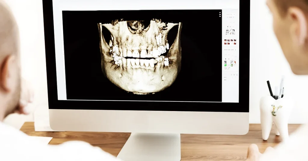
Advanced Imaging with CBCT Technology
Experience precision diagnostic care with the CBCT (Cone Beam Computed Tomography) services at Vlass and Merritt Dental Care in Roswell, GA. Our advanced CBCT technology provides detailed 3D images of your teeth, jaw, and facial bones, aiding in accurate treatment planning and diagnosis. With over 30 years of dental experience, we utilize this cutting-edge imaging to enhance the effectiveness of our treatments, ensuring you receive the most informed and meticulous dental care possible. Trust us to see more clearly and treat more effectively.
Comprehensive Diagnostic Capabilities
With CBCT technology, we can obtain detailed 3D images of the teeth, jawbone, nerves, and surrounding tissues, providing invaluable information for diagnosis and treatment planning. Whether assessing bone density for dental implants or evaluating the extent of dental trauma, our advanced imaging capabilities enable us to identify issues that may not be visible with traditional two-dimensional X-rays.
Precise Implant Planning and Placement
CBCT imaging plays a crucial role in precise implant planning and placement at Vlass & Merritt Dental Care. By visualizing the patient's anatomy in three dimensions, we can accurately determine the optimal location and angle for implant placement, ensuring optimal stability and longevity of the restoration. This precision minimizes the risk of complications and enhances the success rate of dental implant procedures.
Enhanced Treatment Outcomes
Our commitment to utilizing CBCT technology reflects our dedication to providing the highest standard of care and achieving superior treatment outcomes for our patients. With detailed 3D images guiding our treatment decisions, we can customize treatment plans to address each patient's unique needs and goals, leading to more predictable results and greater patient satisfaction.

Advanced Imaging with CBCT Technology
Vlass & Merritt Dental Care utilizes Cone Beam Computed Tomography (CBCT) technology to provide high-resolution 3D images of the oral and maxillofacial structures. This advanced imaging technique allows our skilled dental team to accurately diagnose and plan treatments for a wide range of dental issues, from complex implant placement to TMJ disorders, ensuring precise and effective care for our patients.
CBCT FAQ's
Cone Beam Computed Tomography (CBCT) is a specialized type of medical imaging technology used in dentistry to generate three-dimensional (3D) images of the teeth, jawbones, temporomandibular joints (TMJ), and surrounding structures. It provides detailed, high-resolution images that allow dentists and oral surgeons to visualize the oral and maxillofacial anatomy with precision.
CBCT differs from traditional dental X-rays in several ways. While traditional X-rays produce two-dimensional images, CBCT creates 3D images that provide more comprehensive information about the teeth and jaws, including their spatial relationships and anatomical structures. Additionally, CBCT exposes patients to lower levels of radiation compared to traditional CT scans, making it a safer option for dental imaging.
CBCT is used in various dental specialties, including oral and maxillofacial surgery, orthodontics, periodontics, endodontics, and prosthodontics. It may be utilized to evaluate and plan for dental implants, assess the anatomy of impacted teeth, diagnose temporomandibular joint disorders (TMD), detect and measure pathology such as cysts or tumors, evaluate bone quality and quantity for surgical procedures, and more.
While CBCT involves exposure to ionizing radiation, the amount of radiation used in CBCT imaging is relatively low compared to traditional medical CT scans. Additionally, CBCT scans are performed with a focused beam of radiation that targets the specific area of interest, minimizing radiation exposure to surrounding tissues. Dentists take precautions to ensure that CBCT scans are only performed when necessary and that radiation doses are kept as low as reasonably achievable.
During a CBCT scan, you will be positioned in a stationary unit while a rotating arm takes multiple X-ray images of your head and neck from different angles. The process is painless and typically takes less than a minute to complete. You will be asked to remain still during the scan to ensure clear images. Once the scan is complete, the images are processed by a computer to create detailed 3D reconstructions that can be viewed and analyzed by your dentist.
Read Our 5-Star Ratings
Optimize Your Dental Care with CBCT Imaging
Experience the benefits of advanced CBCT imaging technology at Vlass & Merritt Dental Care. Our skilled dental team harnesses the power of 3D imaging to provide comprehensive diagnostic and treatment solutions tailored to your individual needs. Schedule a consultation today to learn more about how CBCT imaging can enhance your dental care experience.
